Warning
General information only - not dental advice. This guide provides general educational information about dental X-rays. It is not a diagnosis, treatment recommendation, or substitute for professional dental or medical advice. Whether X-rays are appropriate for you depends on your individual clinical situation. Always discuss X-ray decisions with your licensed dentist or oral health provider.
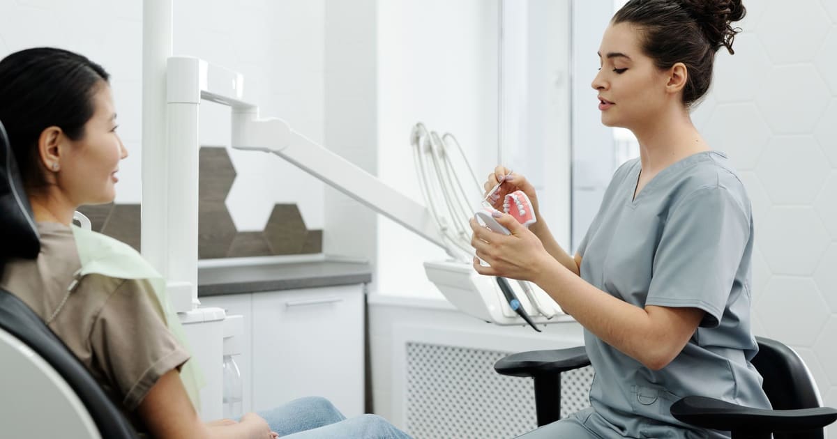
Dental X-rays -- also called radiographs -- allow dentists to detect problems that are not visible to the naked eye, including cavities between teeth, bone loss from gum disease, infections at tooth roots, and the position of unerupted or impacted teeth. The American Dental Association publishes evidence-based guidelines on dental radiograph selection, emphasizing that X-ray recommendations should be individualized based on patient history and risk factors rather than applied on a fixed schedule. This guide explains what the evidence says about frequency, safety, and cost so you can have an informed conversation with your dentist.
Why Are Dental X-Rays Used?
X-rays allow dentists to see structures that a visual examination alone cannot reveal. According to the American Dental Association's clinical guidance, the primary diagnostic purposes of dental radiographs include:
- Detecting cavities (dental caries) between teeth and beneath existing restorations before they are visible or cause symptoms
- Evaluating bone levels around teeth, which is essential for diagnosing and monitoring gum disease
- Identifying infections at tooth roots (periapical pathology) that may not cause obvious symptoms in early stages
- Assessing the position and development of teeth, including wisdom teeth and unerupted teeth in children
- Planning for extractions, implants, orthodontics, and other procedures that require anatomical mapping
Not every dental appointment requires new X-rays. The decision depends on how recently X-rays were taken, whether there have been clinical changes since the last images, and the patient's individual risk profile.
Illustration: four common dental X-ray types and their primary clinical purposes.
How Often Does the ADA Recommend Dental X-Rays?
The American Dental Association published its dental radiograph selection criteria in guidelines available through ADA.org. The guidelines state that there is no universal interval that applies to all patients. Instead, frequency is determined by clinical judgment applied to each patient's individual situation.
For adult patients, the ADA guidelines generally suggest:
- New patients at an initial comprehensive exam: a full-mouth series or panoramic X-ray combined with bitewings, to establish a baseline. The specific recommendation depends on clinical findings during the exam.
- Adult patients with no detected problems and low cavity risk: bitewing X-rays every 24 to 36 months, according to the ADA's radiograph selection criteria table.
- Adult patients with a history of decay or active gum disease: bitewing X-rays may be appropriate more frequently, at intervals determined by the treating dentist.
- Patients who have not been seen by a dentist in several years: a comprehensive baseline set is typically appropriate regardless of risk level.
The ADA guidelines were developed collaboratively with the FDA and are periodically updated based on evolving evidence. Your dentist applies these guidelines in combination with your clinical examination findings.
For guidance on how often to schedule dental visits overall, see our guide on how often to see a dentist.
Are Dental X-Rays Safe? Understanding Radiation Exposure
Radiation exposure is the most common concern patients raise about dental X-rays. The scientific consensus from the American Dental Association, the American College of Radiology (ACR), and the FDA is that modern dental X-rays involve very low radiation doses, and that the clinical benefit of detecting oral disease early typically outweighs the minimal risk associated with this level of exposure.
According to CDC radiation dose comparison data and FDA guidance:
- A single bitewing X-ray delivers a very small effective dose of radiation -- substantially less than what a person receives from background environmental radiation during a typical day
- A full-mouth series (FMX) of approximately 18 images delivers approximately 0.005 millisieverts (mSv) of effective dose
- For comparison, the average US resident receives approximately 3.1 mSv per year from naturally occurring background radiation, according to the CDC
- A chest X-ray delivers approximately 0.1 mSv, and a cross-country commercial flight delivers a similar dose from cosmic radiation
Digital X-ray systems, which have become standard in most US dental offices, use significantly less radiation than older film-based systems while producing higher-resolution images. Lead aprons and thyroid collars further limit exposure to areas not being imaged, and are standard practice in dental offices.
Note
Radiation concerns are reasonable to raise. If you have questions about why a specific X-ray is being recommended or how frequently you have received X-rays, those are reasonable questions for your dentist. The ADA's position is that X-rays should be prescribed only when the clinical benefit -- detecting a problem that changes treatment -- justifies the minimal exposure. You can ask your dentist to explain the clinical rationale for any X-ray before it is taken.
Types of Dental X-Rays and What They Show
Different X-ray types capture different areas and serve different clinical purposes. Understanding the types helps you follow what your dentist is recommending.
Bitewing X-rays are the most common type at routine checkups. Taken with the patient biting down on a small sensor or film tab, they capture the crowns of upper and lower back teeth on the same image. They are the primary tool for detecting cavities between teeth (interproximal decay) and monitoring bone levels for gum disease, according to ADA radiograph selection criteria.
Periapical X-rays show one or two entire teeth from crown to root tip, including the bone surrounding the root. They are used to diagnose infections at the root (periapical abscesses), evaluate root length before root canal treatment, and monitor healing after procedures.
Panoramic X-rays are taken from outside the mouth using a machine that rotates around the patient's head to capture the entire mouth in a single image. They show all teeth, both jaws, the temporomandibular joints, and nearby sinuses. Panoramic X-rays are commonly used to evaluate wisdom teeth, plan implants, or assess jaw problems.
Full-mouth series (FMX) combines periapical and bitewing images covering all teeth. A full-mouth series provides comprehensive baseline information and is commonly taken at a new patient examination or when a complete clinical picture is needed.
Cone beam computed tomography (CBCT) is a three-dimensional imaging system used for complex cases such as implant planning, evaluation of impacted teeth, or assessment of jaw structure. CBCT is not routine and involves higher radiation than standard X-rays. The American Academy of Oral and Maxillofacial Radiology (AAOMR) publishes guidance on when CBCT is clinically appropriate.
How Much Do Dental X-Rays Cost?
X-ray costs vary by type, provider, and geographic market. The table below presents approximate out-of-pocket cost ranges based on published Cigna and Delta Dental cost guides and American Dental Association fee survey data. These are national estimates; actual fees vary significantly by location and practice.
| X-Ray Type | Approximate Out-of-Pocket Range | Common Use |
|---|---|---|
| Single bitewing | $20 - $45 per image | Cavity detection, bone level monitoring |
| Four bitewing set | $80 - $150 | Standard adult checkup |
| Periapical (single) | $25 - $50 per image | Root and surrounding bone |
| Panoramic | $100 - $250 | Full jaw, impacted teeth, jaw problems |
| Full-mouth series | $100 - $300 | Baseline at new patient exam |
Sources: Cigna Healthcare cost guide; Delta Dental cost resources; ADA Health Policy Institute fee survey data. Figures are approximate and vary by region, provider, and year.
Illustration: approximate out-of-pocket cost ranges for common dental X-ray types without insurance. Heights represent range from lower to upper estimate; actual fees vary by region.
Does Insurance Cover Dental X-Rays?
Most dental insurance plans cover diagnostic X-rays as a preventive or basic service, subject to frequency limitations. Delta Dental and Cigna plan documents typically allow:
- Bitewing X-rays: once per calendar year or once every 12 to 24 months, depending on the plan
- Panoramic X-rays: once every three to five years in many standard plans, or when clinically indicated by diagnosis
- Periapical X-rays: often covered as needed for specific diagnostic purposes
- Full-mouth series: typically once every three to five years or at a new patient exam
Coverage specifics depend on your individual plan. If you are unsure whether a recommended X-ray is covered, ask your dental office to verify your benefits before the appointment. Most offices can check your coverage in advance and advise you on your estimated share of the cost.
For context on dental care costs without insurance more broadly, see our guide on cost of dental care without insurance.
Questions to Ask Before Agreeing to X-Rays
Having specific questions ready before X-rays are taken helps you make an informed decision about your care. The Agency for Healthcare Research and Quality (AHRQ) encourages patients to ask about the clinical rationale for any diagnostic test.
Questions worth asking include:
- Why is this X-ray clinically indicated at this visit?
- How long has it been since my last X-rays of this type?
- What problem or condition is this X-ray intended to detect or monitor?
- Is this X-ray covered by my insurance plan at today's visit?
- If not covered, what is the out-of-pocket cost for this type of X-ray?
- Can today's appointment proceed clinically without the X-ray, or is it needed to complete the exam?
Tip
When you transfer to a new dental office, request copies of your most recent X-rays from your previous dentist before your first appointment at the new practice. Most offices will provide them at no charge or for a small administrative fee. Bringing current X-rays can reduce or eliminate the need for repeat imaging, saving both radiation exposure and cost. The ADA supports this practice as part of responsible record transfer between providers.
For a broader look at what to expect and ask during a dental visit, see our guide on questions to ask your dentist.
Frequently asked questions
How often should adults get dental X-rays?
The American Dental Association does not recommend a fixed interval for all adults. Instead, its clinical practice guidelines advise that X-ray frequency should be individualized based on each patient's oral health history, cavity risk, gum disease status, and age. Adults with low cavity risk and stable oral health may need bitewing X-rays every two to three years; higher-risk patients may need them annually, according to ADA guidelines.
Are dental X-rays safe during pregnancy?
The American Dental Association and the American College of Obstetricians and Gynecologists both state that dental X-rays can be performed safely during pregnancy when clinically necessary, particularly with a lead apron and thyroid collar to shield the abdomen. The radiation dose from a standard set of dental X-rays is very low. Discuss your pregnancy with your dentist before any imaging so they can evaluate the clinical need.
How much radiation do dental X-rays expose you to?
According to the American College of Radiology and FDA guidance, a full-mouth series of dental X-rays delivers approximately 0.005 millisieverts (mSv) of effective radiation -- a fraction of the roughly 3.1 mSv of background radiation the average US resident receives annually from natural sources, according to CDC data. A single bitewing X-ray involves substantially less exposure than a full-mouth series.
How much do dental X-rays cost without insurance?
Costs vary by type and region. Based on published Cigna and Delta Dental cost guides, a single bitewing X-ray typically costs $20-$45 per image without insurance; a full-mouth series (FMX) runs roughly $100-$300; a panoramic X-ray (which captures the entire jaw in one image) commonly ranges from $100-$250. These are national estimates and actual fees vary by provider and location.
Does insurance cover routine dental X-rays?
Most dental insurance plans cover routine diagnostic X-rays as a preventive or basic benefit, subject to frequency limitations. Delta Dental and Cigna plan documents typically allow bitewing X-rays once per year or once every two years depending on the plan. A panoramic X-ray may be covered for diagnostic purposes but not automatically at every visit. Check your plan's Explanation of Benefits or call your insurer to confirm your specific allowances.
What is the difference between bitewing and panoramic X-rays?
Bitewing X-rays capture the crowns of upper and lower back teeth in the same image, making them especially useful for detecting cavities between teeth and monitoring bone levels for gum disease. A panoramic X-ray -- taken from outside the mouth with a rotating arm -- captures all teeth, the jawbones, sinuses, and temporomandibular joints in a single image, useful for evaluating impacted teeth, jaw problems, or overall oral anatomy, according to ADA radiograph selection criteria.